
Commentary
By Nithin Jacob
What treatment options are there for individuals whose depression is resistant to anti-depressant drugs? What about the individuals who experience intractable epileptic seizures despite anti-epileptic medication? Or those with schizophrenia who experience debilitating symptoms because they do not respond well to anti-psychotic drugs?
Unfortunately, these situations are not uncommon: medications are not cure-alls. In fact, 1 in 3 patients with depression (1) and epilepsy (2), and 1 in 2 patients with schizophrenia (3), have symptoms that do not respond to drug treatment. Non-drug treatments are emerging as adjunctive therapy for patients with various neurological conditions. One of these promising techniques is transcranial magnetic stimulation (TMS) (2,4).
A safe method for magnetically stimulating the brain, TMS is generally well tolerated by patients (2). How TMS works is by placing an insulated coil on the scalp which then emits a strong, pulsating, magnetic field. Rapid pulses in the magnetic field induce electric currents within the brain (5). This electric current stimulates nearby neurons (i.e., brain cells) to become “activated” (6). The magnetic field does not stimulate the entire brain, however, but just the two to three centimeters of the brain directly beneath the coil (7). Therefore, TMS causes targeted responses in brain cells and without being invasive, unlike surgical techniques.
If this description sounds too heady at the moment, here is an analogy. The mechanisms of TMS work like a puppeteer causing a desired physical movement in the puppet by manipulating the attached strings. Manipulation of the puppet strings represents fluctuations in the magnetic field in TMS. The resulting physical movement of the puppet is analogous to the electrical current produced within the brain’s neurons. When the electrical currents are stimulated in the brain regions responsible for muscle movement, the person’s muscles move as the puppet also does ― without voluntary control.
Through vital developmental phases, TMS has become a therapeutic option for patients with intractable symptoms. First, let’s take a trip back to when TMS was in its infancy before we imagine what its full-fledged future could be like.
Historical background
In the late 1970s, clinicians typically stimulated the brain by sending electricity directly to the scalp (8). This technique was used as a diagnostic tool for patients with multiple sclerosis to evaluate the integrity of the spinal cord and its ability to relay electrical information (9). Although effective, this method often resulted in extreme discomfort, necessitating safer and less painful methods of brain stimulation (8,10).
An encouraging solution was introduced in 1985 by Anthony Barker’s group in England: the world’s first reliable transcranial magnetic stimulator (11). They demonstrated that a single magnetic pulse on the scalp could activate underlying neurons. Clinicians could therefore perform diagnostic tests in patients using this much safer, and far less painful method (10).
Until the late 1980s, TMS was still only used for diagnostic purposes (8). This would change in 1991 when the therapeutic potential of TMS was explored by Alvaro Pascual-Leone in the United States (12). Pascual-Leone and his group discovered that using repetitive transcranial magnetic stimulation (rTMS) not only activated neurons, but also increased the likelihood of future activations. Increasing the chance of a neuron being activated can be referred to as increasing its “excitability.”
Pascual-Leone discovered that these changes in excitability from repeated magnetic pulses lasted even after treatment ended, thus highlighting the therapeutic potential of rTMS (12). For individuals with intractable symptoms, increasing neuronal excitability matters because their brain cells do not respond well or at all to drug treatment ― but may respond to forms of TMS.
With this foundational understanding of TMS’s evolution, let’s investigate the therapeutic uses of rTMS in three clinical conditions: depression, epilepsy, and schizophrenia.

Depression
Approximately 35% of individuals with depression report anti-depressant drug treatment as ineffective (13). Depression is primarily considered a disorder of neuronal activity within two brain networks: the dorsolateral prefrontal cortex (i.e., the region responsible for decision-making) and the ventromedial prefrontal cortex (i.e., the region which helps regulate emotion) (14).
Neurons in the decision-making network have low excitability, meaning that it is difficult to activate these cells (15). In contrast, neurons in the emotional network have higher excitability and are therefore more easily activated (15). This imbalance of neuronal activity results in the persistent low mood associated with depression (14).
Using coordinated magnetic pulses, rTMS may increase excitability in the decision-making network while decreasing excitability in the emotional network ― exerting therapeutic effects on depressive symptoms (14). Pulses made 5-20 times per second enhance neuronal excitability (16), while a pulse delivered once per second has a diminishing effect (17).
A review of 23 studies concluded rTMS treatment significantly improved depressive symptoms compared to a placebo treatment (16). However, the improvement was considered minor because patients receiving rTMS scored only 10% better on clinician-rated depression scales compared to the placebo group. Moreover, these benefits lasted only three to four months post-treatment (16).
Epilepsy
For one third (18,19) of the 50 million people with epilepsy worldwide (20,21), their seizures cannot be controlled through medication. Epilepsy is a neurological disorder that predisposes a person to recurring seizures because of excessive neuronal excitability (22). Sometimes, a brain region prone to this hyperexcitability is the primary motor cortex (23), whose neurons initiate physical movement (24). Uncontrollable activation of these neurons often result in the archetypical body twitching and jerking associated with seizures (5).
Given what you know so far, which frequency of rTMS do you hypothesize would be helpful for those with hyperexcitable neurons? High frequency, or low frequency?
Remarkably, low frequency rTMS in patients with drug-resistant epilepsy can reduce the risk of seizures by 19-35% (25). Applying rTMS with 0.3 and 0.5 pulses per second suppresses the excessive excitability of the neurons, thus reducing the frequency of seizures (26,27).
Researchers believe that the low frequency rTMS is able to provide “relief” for these neurons (5). Think of this like as a water dam. When the water level behind a dam gets too high, there is a risk for the water to overflow. However, releasing some of this water in a controlled manner before it reaches critical levels would reduce the overall water level, and mitigate the risk of an overflow. Controlling the release of water to lower the risk of overflow ― which could cause flooding and destruction downstream ― is analogous to reducing neuronal excitability levels to lower the risk of uncontrollable activation ― which could result in seizures.
Controlling the release of water to lower the risk of overflow ― which could cause flooding and destruction downstream ― is analogous to reducing neuronal excitability levels to lower the risk of uncontrollable activation ― which could result in seizures.
Paradoxically, while rTMS has therapeutic potential in reducing seizures, there is also a small risk of inducing seizures (5,28). Even for an individual without epilepsy, daily exposure to rTMS can potentially lead to seizure attacks (5). Thankfully, rTMS-induced seizures are quite rare and the risk is only concerning when the frequency of magnetic pulses is 60 times per second (29) – the rTMS treatments described above are well below this threshold.
Schizophrenia
Approximately 25-50% of patients with schizophrenia experience symptoms even while on medications (3). Schizophrenia is a psychiatric disorder with two main categories of symptoms: positive and negative. Positive symptoms are defined by new experiences of phenomena such as auditory hallucinations, delusions, and disorganized speech; in contrast, negative symptoms blunt the normal experience of reality, resulting in the inability to feel pleasure, emotional and social withdrawal, and loss of motivational drive (30). While positive symptoms respond well to antipsychotic medication (31), negative symptoms do not (32).
Unfortunately, negative symptoms significantly affect social interactions (e.g., social life at work) and overall quality of life (31). Interestingly, negative symptoms are associated with reduced neuronal excitability in a region of the brain where it is more difficult for neurons to be activated: the left prefrontal cortex (33) (i.e., the region responsible for motivation and decision-making) (34).
Given their reduced neuronal excitability in this brain region, should high frequency or low frequency rTMS be used here?
Yes, high frequency ― and this is why: researchers have tested the effect of rTMS at 8-13 pulses per second on the brain’s motivation and decision-making centre. Compared to a placebo treatment, rTMS significantly reduced the negative symptoms by 30% (31,35), with the effect lasting up to 24 weeks post-treatment (36). At the higher frequency (i.e., 18 pulses per second), negative symptoms were reduced by 38-48% (37).
Returning to the water dam analogy, reduced neuronal excitability can be analogous to water levels that are too low. When the water is below a certain threshold in a hydroelectric dam, it is difficult for electricity to be produced. However, if we were to increase the overall water volume back to the correct level, electricity is easily and efficiently generated. Restoring the optimal water level from low volumes is analogous to increasing neuronal excitability back to a level of normal neuronal function.
Restoring the optimal water level from low volumes is analogous to increasing neuronal excitability back to a level of normal neuronal function.
Reconsidering the questions posed at the start, TMS is an effective treatment option for various clinical populations. As TMS gains momentum in the field of rehabilitation, large-scale studies are needed. Although TMS may not be a “magic pill”, to those individuals with depression, epileptic seizures, and schizophrenia for whom traditional drug therapy is ineffective, it does offer promising clinical benefits.
Acknowledgements
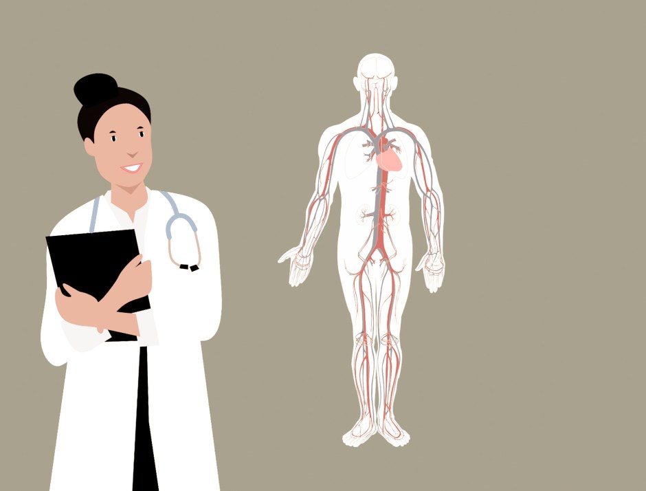
Featured illustration by Ava Schroedl for rehabINK.
Did you enjoy this article? Please help us improve by completing a short reader feedback survey. Thank you!
To refer to this article, it can be cited as:
Jacob N. Transcranial magnetic stimulation (TMS): Magnetic fields and electric currents and brain cells, oh my. rehabINK. 2020;8. Available from: https://rehabinkmag.com
References
- Rush JA, Trivedi MH, Wisniewski SR, Nierenberg AA, Stewart JW, Warden D, et al. Acute and longer-term outcomes in depressed outpatients requiring one or several treatment steps: a STAR*D repor. American Journal of Psychiatry. 2006;163(11):1905–17.
- Pereira LS, Müller VT, da Mota Gomes M, Rotenberg A, Fregni F. Safety of repetitive transcranial magnetic stimulation in patients with epilepsy: A systematic review. Epilepsy and Behavior. 2016;57:167–76.
- Harrow M, Carone BJ, Westermeyer JF. The course of psychosis in early phases of schizophrenia. American Journal of Psychiatry. 1985;142(6):702–7.
- Guo Q, Li C, Wang J. Updated review on the clinical use of repetitive transcranial magnetic stimulation in psychiatric disorders. Neuroscience Bulletin. 2017;33(6):747–56.
- Noohi S, Amirsalari S. History, studies and specific uses of repetitive transcranial magnetic stimulation (rTMS) in treating epilepsy. Iranian Journal of Child Neurology. 2016;10(1):1–8.
- Stuart G, Spruston N, Sakmann B, Häusser M. Action potential initiation and backpropagation in neurons of the mammalian CNS. Trends in Neurosciences. 1997;20(3):125–31.
- Klomjai W, Katz R, Lackmy-Vallée A. Basic principles of transcranial magnetic stimulation (TMS) and repetitive TMS (rTMS). Annals of Physical and Rehabilitation Medicine. 2015;58(4):208–13.
- Horvath JC, Perez JM, Forrow L, Fregni F, Pascual-Leone A. Transcranial magnetic stimulation: A historical evaluation and future prognosis of therapeutically relevant ethical concerns. Journal of Medical Ethics. 2011;37(3):137–43.
- Mills KR, Murray NM, Hess CW. Magnetic and electrical transcranial brain stimulation: Physiological mechanisms and clinical applications. Neurosurgery. 1987;20(1):164–8.
- Green RM, Pascual-Leone A, Wasserman EM. Ethical guidelines for rTMS research. IRB: Ethics and Human Research. 1997;19(2):1–7.
- Barker AT, Jalinous R, Freeston IL. Non-invasive magnetic stimulation of human motor cortex. Lancet. 1985;325(8437):1106–7.
- Pascual-Leone A, Gates JR, Dhuna A. Induction of speech arrest and counting errors with rapid-rate transcranial magnetic stimulation. Neurology. 1991;41(5):697–702.
- Nemeroff CB. Prevalence and management of treatment-resistant depression. Journal of Clinical Psychiatry. 2007;68(suppl 8):17–25.
- Dunlop K, Hanlon CA, Downar J. Noninvasive brain stimulation treatments for addiction and major depression. Annals of the New York Academy of Sciences. 2017;1394(1):31–54.
- Koenigs M, Grafman J. The functional neuroanatomy of depression: Distinct roles for ventromedial and dorsolateral prefrontal cortex. Behavioural Brain Research. 2009;201(2):239–43.
- Health Quality Ontario. Repetitive transcranial magnetic stimulation for treatment-resistant depression: A systematic review and meta-analysis of randomized controlled trials. Ontario Health Technology Assessment Series. 2016;16(5):1–66.
- Latorre A, Rocchi L, Berardelli A, Bhatia KP, Rothwell JC. The use of transcranial magnetic stimulation as a treatment for movement disorders: A critical review. Movement Disorders. 2019;34(6):769–82.
- Kwan P, Brodie MJ. Early identification of refractory epilepsy. New England Joural of Medicine. 2000;342(5):314–9.
- Fisher RS, Cross JH, French JA, Higurashi N, Hirsch E, Jansen FE, et al. Operational classification of seizure types by the International League Against Epilepsy: Position Paper of the ILAE Commission for Classification and Terminology. Epilepsia. 2017;58(4):522–30.
- Kobow K, Blümcke I. Epigenetics in epilepsy. Neuroscience Letters. 2018;667:40–6.
- Banerjee PN, Filippi D, Allen Hauser W. The descriptive epidemiology of epilepsy: A review. Epilepsy Research. 2009;85(1):31–45.
- Fisher RS, Acevedo C, Arzimanoglou A, Bogacz A, Cross JH, Elger CE, et al. ILAE Official Report: A practical clinical definition of epilepsy. Epilepsia. 2014;55(4):475–82.
- Badawy RAB, Strigaro G, Cantello R. TMS, cortical excitability and epilepsy: The clinical impact. Epilepsy Research. 2014;108(2):153–61.
- Shenoy KV, Sahani M, Churchland MM. Cortical control of arm movements: A dynamical systems perspective. Annual Review of Neuroscience. 2013;36(1):337–59.
- Kinoshita M, Ikeda A, Begum T, Yamamoto J, Hitomi T, Shibasaki H. Low-frequency repetitive transcranial magnetic stimulation for seizure suppression in patients with extratemporal lobe epilepsy: A pilot study. Seizure. 2005;14(6):387–92.
- Bae EH, Schrader LM, Machii K, Alonso-Alonso M, Riviello JJ, Pascual-Leone A, et al. Safety and tolerability of repetitive transcranial magnetic stimulation in patients with epilepsy: A review of the literature. Epilepsy and Behavior. 2007;10(4):521–8.
- Menkes DL, Gruenthal M. Slow-frequency repetitive transcranial magnetic stimulation in a patient with focal cortical dysplasia. Epilepsia. 2000;41(2):240–2.
- Misawa S, Kuwabara S, Shibuya K, Mamada K, Hattori T. Low-frequency transcranial magnetic stimulation for epilepsia partialis continua due to cortical dysplasia. Journal of the Neurological Sciences. 2005;234(1–2):37–9.
- George MS, Lisanby SH, Sackeim HA. Transcranial magnetic stimulation: Applications in neuropsychiatry. Archives of General Psychiatry. 1999;56(4):300–11.
- Liu J, Jia J, Chua X, Chong SA. The impact of emotion dysregulation on positive and negative symptoms in schizophrenia spectrum disorders: A systematic review. 2020;1–13.
- Prikryl R, Kasparek T, Skotakova S, Ustohal L, Kucerova H, Ceskova E. Treatment of negative symptoms of schizophrenia using repetitive transcranial magnetic stimulation in a double-blind, randomized controlled study. Schizophrenia Research. 2007;95(1–3):151–7.
- Kirkpatrick B, Fenton WS, Carpenter WT, Marder SR. The NIMH-MATRICS consensus statement on negative symptoms. Schizophrenia Bulletin. 2006;32(2):214–9.
- Hill K, Mann L, Laws KR, Stephenson CME, Nimmo-Smith, McKenna PJ. Hypofrontality in schizophrenia: A meta-analysis of functional imaging studies. Acta Psychiatrica Scandinavica. 2004;110(4):243–56.
- Strafella AP, Paus T, Barrett J, Dagher A. Repetitive transcranial magnetic stimulation of the human prefrontal cortex induces dopamine release in the caudate nucleus. Journal of Neuroscience. 2001;21(15):1–4.
- Jin Y, Potkin SG, Kemp AS, Huerta ST, Alva G, Thai TM, et al. Therapeutic effects of individualized alpha frequency transcranial magnetic stimulation (αTMS) on the negative symptoms of schizophrenia. Schizophrenia Bulletin. 2006;32(3):556–61.
- Quan WX, Zhu XL, Qiao H, Zhang WF, Tan SP, Zhou DF, et al. The effects of high-frequency repetitive transcranial magnetic stimulation (rTMS) on negative symptoms of schizophrenia and the follow-up study. Neuroscience Letters. 2015;584:197–201.
- Linsambarth S, Jeria A, Avirame K, Todder D, Riquelme R, Stehberg J. Deep transcranial magnetic stimulation for the treatment of negative symptoms in schizophrenia: Beyond an antidepressant effect. Journal of ECT. 2019;35(4):46–54.
