Original Research & Commentary
By Vahid Anwari, Behruz Karasfi, & Ashley Lai
Imagine you had the power to make any object in the world. What would you create first?
Today, your dream is nearly reality through technology called three-dimensional (3D) printing, or rapid prototyping. The applications of this technology are extremely diverse. For example, a company can use it to make parts for an aircraft or a prosthetic nose for a burn victim. 3D printing refers to the production of a physical object from a digital image. The process typically involves depositing and fusing layers of materials by a 3D printer.
There are many types of 3D printers, but the most common are material jetting and powder bed fusion. Material jetting is often used for medical models because of its versatility. It can print using photopolymers, which are resins that harden when exposed to ultra-violet light. Photopolymers can assume a wide range of properties—rigid or flexible, transparent or opaque—allowing for the integration of multiple colours and material properties in a single model. Powder bed fusion printing uses a fine powder to build a model one layer at a time. It is used to 3D print medical devices and implants using thermoplastics or metals, such as nylon or titanium alloy.
3D printing is especially useful in the design and development of custom-tailored implants and personalized prostheses for patients undergoing rehabilitative therapy. The major advantage of 3D printing over traditional methods is that several models can be created virtually using 3D rendering software prior to the final model being printed in-house. This advantage facilitates the role of 3D printing in customizing healthcare for patient-centred precision medicine.
The benefits and applications of 3D printed models (3DPMs) in medicine have been demonstrated in medical education and surgical planning. However, the benefits and uses of 3DPMs in rehabilitation sciences has not yet received similar consideration. To explore the future of this novel technology, we describe the fundamentals of 3D printing methodology and highlight the enormous potential for 3DPMs in rehabilitation.
Can I print a 3D model in my own garage?
Absolutely! Step one of the 3D printing process involves visualizing the model, choosing the appropriate materials, and deciding on a suitable type of 3D printer. Building an accurate model for medical applications requires the assistance of professionals from various disciplines. In the rehabilitation context, input might be required from a multi-disciplinary team including radiologists, technologists, physicists, biomedical engineers, and/or rehabilitation professionals (e.g., physical or occupational therapists).
The desired goals are identified, including characterizing the material properties of the target body tissues (e.g., heart tissue differs from lung tissue). Next, optimal 3D printing materials will need to be selected—a decision largely dependent on the anatomical structures of interest. Finally, the 3D printer is selected from the wide range of commercially available printers. The choice of printer depends on factors including financial constraints, the estimated volume usage, and the desired resolution of the final 3DPM.
Step two of the 3D printing process is to capture accurate images of the desired model. For anatomical models (e.g., an upper limb), the image must be a format known as Digital Imaging and Communications in Medicine (DICOM) that contains information such as volume, density, and thickness. Images in DICOM format can be captured using computed tomography (CT), magnetic resonance (MR), or ultrasound, before being converted to 3D print files. The most used images are from CT due to their high contrast, signal-to-noise ratio, and spatial resolution. Together, these characteristics improve the differentiation of structures and facilitate image post-processing.
Step three of the 3D printing process is to digitally isolate the anatomy that is to be printed from the entire image set. Using the images acquired from CT or MR, a medical imaging technologist carefully isolates the anatomical structures of interest. Commercially available or free, open-source software can be used to complete this step (1,2).
Figure 1 shows an example of a model of the trachea (i.e., windpipe) and the lungs isolated using advanced digital visualization software. This model can be used to print the entire trachea for a recipient, or parts thereof. Typically, the process of building a virtual model from images takes anywhere from 30 minutes to three hours, depending on the complexity of the structures

Am I ready to print the model yet?
Almost! Step Four in the 3D printing process is to clean and prepare the final model for printing. This step is necessary to ensure the model is printed with the desired resolution and structural integrity. The process consists of manually closing open gaps in the file, smoothing out surfaces, and adding support structures. Once the model has been finalized, the file is converted to a readable format.
Figure 2 displays a snapshot of the steps necessary to 3D print a prosthetic foot. In this case, 3D visualization software (i.e., Slicer) was used to manipulate the anatomical model.

Step Five, the last step in the process, is the 3D printing itself, which can take an hour to over 24 hours for larger models. The 3D printer builds up layers of the chosen material according to a programmed pathway as the object takes shape on the printing bed. Figure 3 shows a completed 3DPM of the liver.

So 3D printing has enormous potential in rehabilitation sciences?
Definitely! One of the most exciting uses of 3D printing in rehabilitation sciences is developing personalized prosthetics using a patient’s own medical images, and at low cost. For example, a prosthetic arm takes approximately six hours to print and costs about $100 using a consumer-grade printer (3). At a price tag equivalent to a months’ worth of Starbucks coffee, a 3D printed prosthetic limb can provide substantial improvements in functionality for patients much more quickly and affordably than traditional models currently used in healthcare centres around the world.
Patients that can benefit from 3DPMs include individuals requiring amputation due to diabetes or gangrene, and those with severe musculoskeletal injuries or burns. For example, Lee and colleagues (4) showed that laboratory-made skin models can be printed using an eight-channel droplet-based printer that inserts skin cells and structural cells into a matrix, creating an accurate representation of the dermal (i.e., deep) and epidermal (i.e., superficial) skin layers. In this case, the outstanding challenge is to develop blood vessels for nutrient and oxygen delivery and waste removal in the printed tissues.
One of the most exciting uses of 3D printing in rehabilitation sciences is developing personalized prosthetics using a patient’s own medical images, and at low cost.
Another exciting opportunity for 3D printing is the development of exoskeletons (i.e., devices worn outside the body) for physical therapy. These devices have been used in clinical and home-based rehabilitation settings to address hand impairments such as spasticity or muscle weakness resulting from stroke, paralysis, or neuromuscular injury. 3D printing methodology can be used to design and build anatomically accurate hand prostheses that can be controlled by the user. Other 3D exoskeleton devices created for patients with neuromuscular impairments include prosthetic lower legs, arms, feet, and hands.
For aesthetic reconstruction, specific sections of the face, nose, and ear can be printed using fused filament fabrication (i.e., heated filament of thermoplastic built up in continuous layers) or stereolithography (i.e., liquid polymer hardened by a computer-controlled laser beam). Both techniques can create highly detailed molds that can be cast in silicone to create a natural appearance (5).
One of the most common types of lower limb devices are ankle-foot orthoses (AFOs). The traditional method requires an orthotist to build a positive mold of the ankle and foot which is then used to shape the AFO from thermoplastic. The entire process typically takes eight days, including supplier manufacturing. In contrast, 3D printing of an AFO can be completed in two days, including delivery to the recipient. Telfer and colleagues (6) successfully demonstrated an “adjustable stiffness AFO” that allowed the user to modify the device according to their needs. For example, a stiffer device is suitable for walking on flat surfaces while a more flexible device is desirable for climbing stairs.
Additional developments in assistive technology using 3D printing include the development of tactile displays of graphs for people with visual impairments. Medola and colleagues (7) used 3D printing to develop a new type of pushrim for wheelchairs and recruited six participants to test the new design. Participants were satisfied with the 3D-printed pushrim, reporting that it enabled easier maneuvering of the wheelchair in tight spaces (7).
But there are the limitations going forward, right?
Right. Currently, the literature reports several diverse applications for 3DPMs in rehabilitation sciences. The main advantage is that it allows for accurate development of customizable and precise anatomical models; however, there are no standardized measures for assessing the performance of 3DPMs. Accordingly, there are limits to the breadth and quality of research on using 3DPMs for rehabilitation.
One reason that 3D printing has not gained traction in rehabilitation sciences is that a successful 3D printing project demands collaboration between researchers and clinicians who may not otherwise cross paths (8). For example, many researchers in biomechanical engineering and medical imaging departments are well-versed in 3D printing but may not be familiar with current research and clinical practice in rehabilitation. Collaboration between professionals in these fields could yield enormous benefits for patients.
After all this, do you still want to print an accurate anatomical model in your own garage? If so, here’s how you get started: the first step is to conceptualize! The National Institutes of Health contains a vast repository of free, downloadable images of anatomical models that you can use to start printing your own models (9).
May we recommend focusing on rehabilitation sciences?
Acknowledgements
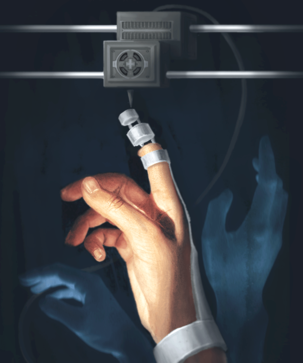
Featured illustration by Ren YuXiang for rehabINK.
To refer to this article, it can be cited as:
Anwari V, Karasfi B, & Lai A. 3D printing for rehabilitation sciences: Are we there yet?. rehabINK. 2020;8. Available from: https://rehabinkmag.com
References
- 3D Slicer [Internet]. [cited 2019 Dec 8]. Available from: https://www.slicer.org/
- Fedorov A, Beichel R, Kalpathy-Cramer J, Finet J, Fillion-Robin JC, Pujol S, et al. 3D Slicer as an image computing platform for the Quantitative Imaging Network. Magnetic Resonance Imaging. 2012;30(9):1323–41.
- Ibrahim AMS, Jose RR, Rabie AN, Gerstle TL, Lee BT, Lin SJ. Three-dimensional printing in developing countries. Plastic and Reconstructive Surgery Global Open. 2015;3(7):e443.
- Lee V, Singh G, Trasatti JP, Bjornsson C, Xu X, Tran TN, et al. Design and fabrication of human skin by three-dimensional bioprinting. Tissue Engineering Part C Methods. 2014;20(6):473–84.
- Liaw CY, Guvendiren M. Current and emerging applications of 3D printing in medicine. Biofabrication. 2017;9(2):024102.
- Telfer S, Pallari J, Munguia J, Dalgarno K, McGeough M, Woodburn J. Embracing additive manufacture: Implications for foot and ankle orthosis design. BMC Musculoskeletal Disorders. 2012;13(1):84.
- Lunsford C, Grindle G, Salatin B, Dicianno BE. Innovations with 3-dimensional printing in physical medicine and rehabilitation: A review of the literature. Physical Medicine and Rehabilitation. 2016;8(12):1201–12.
- Chepelev L, Wake N, Ryan J, Althobaity W, Gupta A, Arribas E, et al. Radiological Society of North America (RSNA) 3D printing Special Interest Group (SIG): Guidelines for medical 3D printing and appropriateness for clinical scenarios. 3D Printing in Medicine. 2018;4(1):11.
- NIH 3D Print Exchange [Internet]. [cited 2019 Dec 9]. Available from: https://3dprint.nih.gov/discover/3dpx-000906
